Nerve Root MRI

22. September 2021
Author:s
Dr. med.
Michael Ho
Certified radiology specialist
Expert for Peripheral Nerves
Table of contents

What is a Nerve root MRI?

With a 3D Nerve root MRI at ARISTRA, we could help a patient determine the exact location of his sustained injuries. Find out how PD Dr. med. Daniel Truhn managed to help with this vital step on the patient's way to recovery.

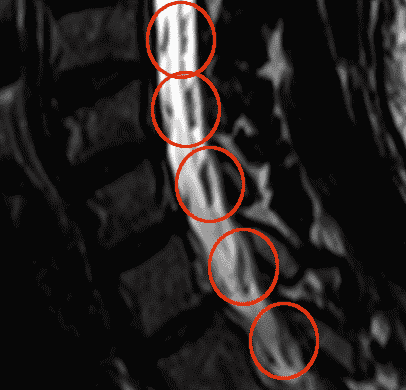
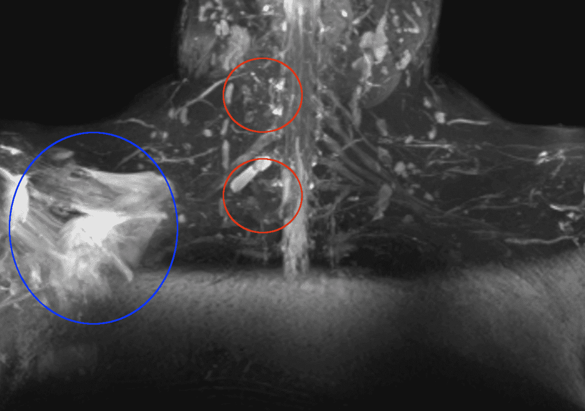
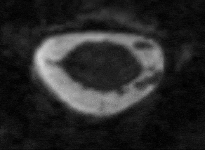
The patient was referred to us after having suffered severe injuries in a traffic accident. Movement in the right arm was mostly lost. To determine how to progress with the treatment, it was of utmost importance to determine the exact site of the injury (www.thenerve.net/journal/view.php?number=90):
  • If the injury is postganglionic (i.e. if the nerve is ruptured after having passed the first “relay station” close to the spinal cord), nerve grafting is possible which is considered the best surgical option and which in principle allows for full restoration of the function.
  • If the injury is preganglionic (i.e. in the early course of the nerve close to the spinal cord), nerve grafting is not an option. Some surgical options remain, but the outcome is usually worse.

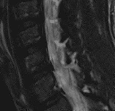
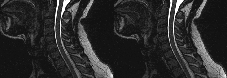
The patient had initially undergone an MRI examination at a major university hospital in Germany, but the exact site of the injury could not be determined due to insufficient image quality.
We worked together with imaging specialists to optimize image quality for the examination.

Optimization of Image Quality and Choice of Sequence

Due to the small size of the spinal nerves, high image resolution is paramount. Conventional two-dimensional image acquisition in which the MR images are acquired slice wise is not suitable to deliver the high resolution. Three-dimensional image acquisition on the other hand is capable of generating images with high resolution but suffers from long scan times and potentially low signal. To remedy these shortcomings, we chose a special technique called constructive interference in steady state (CISS), which restores signal intensity and allows for fast image acquisition over a small area. CISS exhibits a high signal for the spinal fluid and a low signal for the nerve, thus the nerve appears dark against a bright background.
Nerve root MRI conventional 2D series
Conventional 2D series
Nerve root MRI conventional 3D series
3D series, the nerve roots are clearly distinguishable (red circles)

Acquisition of Images

The total imaging session in the MRI took about 50 minutes. No contrast agent was necessary to depict the nerves accurately. We acquired conventional two-dimensional images to depict the anatomy and plan the higher resolution three-dimensional sequences.
Shoulder MRI with Nerve root damage
Fat suppressed sequence giving a good overview and demonstrating the nerve damage (red circles) and the injuries of the shoulder muscles (blue circle).

Diagnostic Findings

3D sequences offer superior resolution depicting the nerve roots which are partially missing on in the right series (right patient side) and are present on the left series.
Nerve root MRI 3D sequences
Based on the high-resolution 3D images, we could clearly identify the injury of the nerve roots at different levels (C5, C6, C8, Th1) establishing a preganglionic injury. The fine nerve roots are best visualized on the axial slices as shown in the following series:
Nerve root MRI of the spinal cord
The video shows an axial view of the spinal cord (dark area in the center) within the surrounding liquor (bright area) on two neighbouring heights. While both nerve roots are intact on one level, the attachment of the right nerve root (note that it appears on the left image side) is torn and missing.

Conclusion

Nerve root injuries can be depicted with high resolution MRI. It is best to employ a real three-dimensional acquisition scheme (as opposed to the much wider used two-dimensional acquisition) in order to have equal resolution in all directions (so called isotropic resolution). This allows accurate assessment of the attachment of the nerve roots to the spinal cord.
Book your appointment with ARISTRA

Make MRI appointment

You can use our convenient online form. We will get back to you as soon as possible. We can usually offer you an appointment for your MRI at short notice.
EN Blog